Ultrasound-Ventricular Shape Analysis in Prediction of Hemorrhagic Hydrocephalus in Premature Neonates
Need:
Premature neonates with intraventricular hemorrhage followed by hydrocephalus (post-hemorrhagic hydrocephalus, PHH) are at the highest risk of severe palsy and other adverse neurodevelopmental outcomes.
Solution:
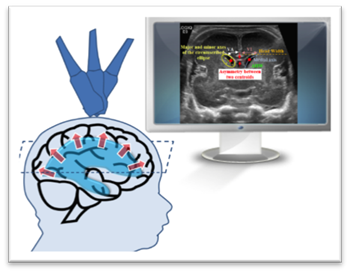
Our team of neurologists, neonatologists, radiologists and quantitative imaging specialists is developing a new quantitative imaging framework to automatically quantify the morphology of the cerebral ventricles in early CUS. This solution incorporates advanced quantitative imaging and machine learning methods to analyze the morphological features in order to predict the potential need for temporizing intervention in premature neonates. The proposed tool allowed us to establish a quantitative method for PHH evaluation on CUS in extremely premature neonates with intraventricular hemorrhage.
Impact:
Our tool would be useful for neurosurgical consultation teams (neurology, neonatology and neurosurgery) to make a decision early without any bias in a safe way. Also, it could help identify neonates who are at risk sooner and decide early preventive treatment.
Partners:
- Division of Neurology
- Division of Neonatology
- Beaumont Hospital
Funding:
Thrasher Research Fund